Rib Cage Anatomy Labeled / 3d Illustration Of Human Skeleton System Rib Cage With Labels Anatomy Anterior View Canstock : Learn vocabulary, terms, and more with flashcards, games, and other study tools.
Rib Cage Anatomy Labeled / 3d Illustration Of Human Skeleton System Rib Cage With Labels Anatomy Anterior View Canstock : Learn vocabulary, terms, and more with flashcards, games, and other study tools.. The front of the structure is the sternum, which is commonly called the breast bone. Start studying rib cage anatomy. 16 photos of the rib cage diagram with organs. The rib cage is a structure comprised of numerous bones and cartilage. The thoracic cage (rib cage) forms the thorax (chest) portion of the body.
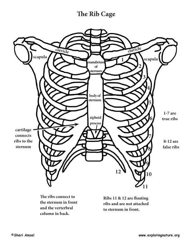
From the back, the ribs angle down slightly. The rib cage labeled diagram. Anatomy of the rib cage diagram in this image, you will find thoracic vertebrum, costochondral joint, costal cartilage, costal margin, costal arch, thoracic vertebrum, xiphoid process, xiphisternal joint, body, manubrial sternal joint, manubrium, the sternal notch in it. In the back, each rib attaches into the thoracic vertebrae. Seer training axial skeleton 80 bones.
Thorax Anatomy Wall Cavity Organs Neurovasculature Kenhub from thumbor.kenhub.com Numbered ribs, sternum, cartilage parts and clavicular articulation. Its functions are to protect the thoracic organs from trauma and also form the bony attachment for various muscles. Lumbar spine diagram labeled wiring diagram t1. Rib cage anatomy, labeled vector illustration diagram. Rib bones are not classified as long bones.instead, anatomists classify the ribs as flat bones, and they are located within the axial skeleton.together with the sternum, thoracic vertebrae, and costal cartilages, the ribs form the thoracic cage, also called the bony thorax. From the back, the ribs angle down slightly. The thoracic cage (rib cage) forms the thorax (chest) portion of the body. Lateral view of a pair of ribs articulating with the thoracic vertebrae.
Click on the tags below to find other quizzes on the same subject.
Rib cage anatomy, labeled vector illustration diagram. Vector steak meat hand drawing with pepper and rosemary. There is costal cartilage that bridges the ribs and the sternum together. This is a preview video for our tutorial about the anatomy of the ribs, the different types, their. 16 photos of the rib cage diagram with organs. The upper edge is round and the lower sharp. Medical human chest skeletal bone structure model. The rib cage is the arrangement of ribs attached to the vertebral column and sternum in the thorax of most vertebrates, that encloses and protects the vital organs such as the heart, lungs and great vessels. Rib cage anatomy, labeled vector illustration diagram. Start studying rib cage anatomy. They also have a role in ventilation; In the back, each rib attaches into the thoracic vertebrae. The average skeleton contains 24 individual ribs, formed in 12.
The rib cage is a structure comprised of numerous bones and cartilage. Its functions are to protect the thoracic organs from trauma and also form the bony attachment for various muscles. There is a printable worksheet available for download here so you can take the quiz with pen and paper. In the back, each rib attaches into the thoracic vertebrae. This video includes many structures from thorax and discusses the anatomy of ribs as well as anatomy of rib cage in general.
Shoulder Rib Cage And Upper Limb from www.exploringnature.org The rib cage is a primarily protective structure, encircling the heart and lungs. Rib bones are not classified as long bones.instead, anatomists classify the ribs as flat bones, and they are located within the axial skeleton.together with the sternum, thoracic vertebrae, and costal cartilages, the ribs form the thoracic cage, also called the bony thorax. The ribs are a set of twelve paired bones which form the protective 'cage' of the thorax. Our latest youtube film is ready to run. Its functions are to protect the thoracic organs from trauma and also form the bony attachment for various muscles. Learn vocabulary, terms, and more with flashcards, games, and other study tools. 16 photos of the rib cage diagram with organs. Anatomical ribs 12 photos of the anatomical ribs anatomical name for floating ribs, anatomical term ribs, anatomical word for ribs, anatomy.
They also have a role in ventilation;
In the back, each rib attaches into the thoracic vertebrae. There are twelve (12) pairs of ribs and all articulate posteriorly with the thoracic vertebrae. Numbered ribs, sternum, cartilage parts and clavicular articulation. An uneven rib cage means the two sides of the rib cage are not symmetrical. Medical human chest skeletal bone structure model. There is a printable worksheet available for download here so you can take the quiz with pen and paper. They articulate with the vertebral column posteriorly, and terminate anteriorly as cartilage (known as costal cartilage). The front of the structure is the sternum, which is commonly called the breast bone. Rib cage anatomy labeled vector illustration diagram stock vector. Anatomy of the thoracic skeleton ribcage medical chart. 16 photos of the rib cage diagram with organs. On the interior wall of the rib body is a channel, sulcus costae, with blood vessels and nerves. Lumbar spine diagram labeled wiring diagram t1.
Numbered ribs, sternum, cartilage parts and clavicular articulation. The upper edge is round and the lower sharp. Rib cage, in vertebrate anatomy, basketlike skeletal structure that forms the chest, or thorax, and is made up of the ribs and their corresponding attachments to the sternum (breastbone) and the vertebral column. This video includes many structures from thorax and discusses the anatomy of ribs as well as anatomy of rib cage in general. They also have a role in ventilation;
Rib Cage Wikipedia from upload.wikimedia.org There are twelve (12) pairs of ribs and all articulate posteriorly with the thoracic vertebrae. The front of the structure is the sternum, which is commonly called the breast bone. Seer training axial skeleton 80 bones. Anatomy of the rib cage diagram in this image, you will find thoracic vertebrum, costochondral joint, costal cartilage, costal margin, costal arch, thoracic vertebrum, xiphoid process, xiphisternal joint, body, manubrial sternal joint, manubrium, the sternal notch in it. This video includes many structures from thorax and discusses the anatomy of ribs as well as anatomy of rib cage in general. Rib bones are not classified as long bones.instead, anatomists classify the ribs as flat bones, and they are located within the axial skeleton.together with the sternum, thoracic vertebrae, and costal cartilages, the ribs form the thoracic cage, also called the bony thorax. Learn vocabulary, terms, and more with flashcards, games, and other study tools. Start studying rib cage labeling.
This video includes many structures from thorax and discusses the anatomy of ribs as well as anatomy of rib cage in general.
| find, read and cite all the research you need on researchgate. Rib cage anatomy, labeled vector illustration diagram. From the back, the ribs angle down slightly. It also supports the shoulders and upper limbs. There is costal cartilage that bridges the ribs and the sternum together. Of all 24 ribs, the first seven pairs are often labeled as 'true.' these bones are. The labels displayed in black are originals whereas the labels displayed in red colour are mirror images. Check out our articles, video tutorials, quizzes, and labeled diagrams. The upper edge is round and the lower sharp. The average skeleton contains 24 individual ribs, formed in 12. An uneven rib cage means the two sides of the rib cage are not symmetrical. The thoracic cage protects the heart and lungs. Related posts of rib cage diagram labeled anatomical diagram of internal organs.


0 Komentar